Sunday, January 12, 2014
Monday, January 6, 2014
The Heart
The human heart has four chambers and they are the right atrium, left atrium, right ventricle and left ventricle. The septum is a muscular wall that separates the right side of the heart from the left. The right side of the heart pumps deO2 blood to the lungs and the right side of the heart pumps O2 blood to the body systems. The atria are the receiving chambers, the right atrium receives deO2 blood from body systems via the superior and inferior vena cava, the left atrium receives O2 blood from the lungs via the pulmonary veins. The ventricles are sending chambers. The right ventricle sends deO2 blood to the lungs through the pulmonary valve, pulmonary trunk and the pulmonary arteries. The left ventricle sends O2 blood to the body systems through the aortic valve and aortic arch or dorsal aorta.

The heart has other things that make it works, for example, Atrioventricular valves separate the atria from the ventricle. There are two different types of AV valves and they are tricuspid valve which is on the right side and the bicuspid valve which is on the left side. They open when the atria contract and close when the ventricle contracts to prevent backflow. The chordae tendinae are tendon-like pieces of tissue, they keep the AV valves from inverting when the ventricles contract. Papillary muscle holds and supports the chordae tendinae.

The SA node if found along the wall of the upper right atrium chamber, it stimulates the simultaneous contraction of the atria and sends a nerve impulse to the AV node. The SA node initiates the heartbeat, this is why it has a nickname of being the pacemaker. The AV node is in the lower right atrium close to the AV valve. When the AV node receives the nerve impulse from the SA node it fires to initiate the contraction of the large ventricles by sending the message through the purkinje fibres. All of this is what moves our blood around our body and it is what we feel when we feel the pulse or listen to the heartbeat.

EKG (Electrocardiogram)
This is a machine the measures the systolic and diastolic of the heart. It is measured and is represented by the letter P,Q,R,S and T. The P represents the simultaneous contraction of the atria (caused by the SA node), the QRS is the contraction of the ventricles (caused by the AV node and purkinje fibres) and the T is the recovery of the ventricles.

The heart has other things that make it works, for example, Atrioventricular valves separate the atria from the ventricle. There are two different types of AV valves and they are tricuspid valve which is on the right side and the bicuspid valve which is on the left side. They open when the atria contract and close when the ventricle contracts to prevent backflow. The chordae tendinae are tendon-like pieces of tissue, they keep the AV valves from inverting when the ventricles contract. Papillary muscle holds and supports the chordae tendinae.
The SA node if found along the wall of the upper right atrium chamber, it stimulates the simultaneous contraction of the atria and sends a nerve impulse to the AV node. The SA node initiates the heartbeat, this is why it has a nickname of being the pacemaker. The AV node is in the lower right atrium close to the AV valve. When the AV node receives the nerve impulse from the SA node it fires to initiate the contraction of the large ventricles by sending the message through the purkinje fibres. All of this is what moves our blood around our body and it is what we feel when we feel the pulse or listen to the heartbeat.
EKG (Electrocardiogram)
This is a machine the measures the systolic and diastolic of the heart. It is measured and is represented by the letter P,Q,R,S and T. The P represents the simultaneous contraction of the atria (caused by the SA node), the QRS is the contraction of the ventricles (caused by the AV node and purkinje fibres) and the T is the recovery of the ventricles.
P= atrial systole (contract)
R= ventricular systole (contract)
S= atrial diastole (rest)
T= ventricular diastole (rest)
HOW DOES THE HEART BEAT ON ITS OWN?
The regulation of the heartbeat is under the influence of the autonomic nerve system. The part of the brain that governs the speed of the heart rate is called the medulla oblongata. It will speed up or down the heart rate when needed. Under normal circumstances, the heart controls itself. This is why the heart can still beat 2-5 min after it is removed from the body.
BLOOD PRESSURE
High blood pressure (hypertension) puts a constant strain on the tissues and it may cause capillaries to burst. If this happens in the brains it is a stroke, if it happens in the heart then it is a heart attack. The longer a person has high blood pressure, the greater the potential for tissue damage. Sometimes high blood pressure is normal for example when doing physical activities, but the brain should return the blood pressure back to normal (lower level). Low blood pressure (hypotension) is particularly not a good thing to have either. It can result from genetics, anemia, dehydration, blood loss and shock. Proper kidney function can only be maintained if there is a sufficient pressure for filtration. Some factors that can affect blood pressure can include:
1. Vessel Diameter (bigger=lower BP, higher=higher BP)
2. Blood viscosity (thick= higher BP, thin = lower BP)
3. Total Blood Vessel Length (more fat = higher BP, thinner = lower BP)
4. Cardiac output (faster heartbeat = higher BP, slower heartbeat = lower BP)
5. Stress (lots = higher BP, less = lower BP)
Circulatory system
In the Circulatory system there are five types of blood vessels and they are:
1. Arteries: Transport blood away from the heart. They have thick elastic walls so that the walls can expand when it needs too. The expansion of the arteries is the pulse we feel. Arteries are usually found deep and along bones in the body. This protects them from injury and temperature loss. Arteries have a very high blood pressure.

2. Arterioles: Control the blood flow to capillaries. They are smaller in diameter than arteries, they have thinner walls. They also have a pre-capillary sphincter, they contract and dilate to increases or decrease blood flow to a particular capillary bed. Arterioles are located leading towards all capillaries. Blood pressure > osmotic pressure.


Capillary Fluid Exchange
Net blood pressure forces water out of the blood into the interstitial fluid. The water carries O2 and nutrients with is out. Because there is more O2 and nutrients in interstitial fluid, it diffuses into body cells. The large things for example RBC, WBC, platelets and blood proteins stay in the capillary because they are too big to leave. Since most of the water has left, the blood becomes very concentrated. The venule side of the capillary is therefore under great osmotic pressure to draw back water into the blood. The water carries CO2 and metabolic wastes(urea), these are carried to the kidneys and other excretory organs to be removed.

1. Arteries: Transport blood away from the heart. They have thick elastic walls so that the walls can expand when it needs too. The expansion of the arteries is the pulse we feel. Arteries are usually found deep and along bones in the body. This protects them from injury and temperature loss. Arteries have a very high blood pressure.
2. Arterioles: Control the blood flow to capillaries. They are smaller in diameter than arteries, they have thinner walls. They also have a pre-capillary sphincter, they contract and dilate to increases or decrease blood flow to a particular capillary bed. Arterioles are located leading towards all capillaries. Blood pressure > osmotic pressure.
3. Capillaries: Connect arteries to veins and are the site of the capillary fluid exchange. They have very thin walls and are found everywhere within a few cells of each other.
4. Venules: They drain blood from the capillaries and join to form veins. They have thinner walls then veins. The are located often near the surface. Osmotic pressure > Blood pressure.
5. Veins : Transport blood towards the heart. Veins have inelastic walls that contains valves that prevent blood from flowing backwards.They are often near the surface and are surrounded by skeletal muscle. 'squeeze' the blood along. Blood pressure and velocity is much lower than in arteries.
Some major arteries and veins are:
1. Subclavian arteries bring blood to the chest wall and arms. Subclavian veins bring blood away from the chest wall and arms to the superior vena cava of the heart.
2.Carotid arteries bring blood to the brain. Jugular veins carry blood away from the brain to the superior vena cava of the heart.
3. Mesenteric arteries bring blood to the digestive system. Then the hepatic portal vein carries the blood from the digestive system to the liver. The Hepatic vein takes the blood from the liver to the inferior vena cava of the heart. The Hepatic arteries tale blood from the heart to the liver.
4. Renal arteries take O2 blood to the kidneys and the renal veins takes the deO2 blood from the kidneys to the inferior vena cava of the heart.
5. Iliac arteries take O2 blood from the heart to the trunk and legs and the iliac veins takes the deO2 blood from the trunk and legs to the inferior vena cava of the heart.
6. The coronary Arteries brings O2 blood to the heart and the coronary vein takes deO2 blood from the heart to the inferior vena cava.
7. The Pulmonary arteries take deO2 blood away from the heart to the lungs to get replenished with O2. The Pulmonary veins take O2 rich blood from the lungs into the heart where that blood will be sent out to the rest of the body systems.
The aorta take O2 blood to the arteries for the top part of the body the dorsal aorta takes O2 blood to the arteries involving the lower part of the body.
Systemic circulation is the system of blood vessels that delivers O2 blood to the body systems. Pulmonary circulation is the system of blood vessels that delivers deO2 blood to the lungs to be replenished with oxygen.
How to do a blood trace: To do a blood trace you follow the way the blood goes through the body and name all the parts it goes though. For example:
Something that can help you do this if you don't have the circulatory system memorized is this:  However, it is better that one memorizes the system.
However, it is better that one memorizes the system.
Fetal Circulation
A fetus does not use its lungs, it receives O2 blood from the placenta. There are four features that fetus' have that adults do not and they are:
Foramen Ovale: This is an opening between the left and right atria, it is covered by a flap that acts as a valve. It allows the blood to by pass the lungs and reroutes most of the blood from the right atrium into the left atrium.
Ductus Arteriosus (Arterial Duct): This is a small arterial connection, like a shunt, between the pulmonary artery and the aorta. It further allows blood to bypass the lungs.
Umbilical Cord: Has three blood vessels traveling through it. The largest one is the umbilical vein, which transports blood with oxygen and nutrients to the fetus. The other two are the umbilical arteries, which branch off of the iliac arteries in the fetus and take used (CO2 and wastes) blood back into the mother via the placenta.
Ductus Venosus: This blood vessel connects the umbilical vein to the vena cava. The O2 blood from the umbilical vein mixes with deO2 blood in the vena cava. The duct venosus bypasses the liver and this blood is sent directly to the heart, blood will eventually go to the liver but not until it reached the hepatic portal vein. This is why the fetus is so susceptible to toxins in the blood.
Lymphatic System
The lymphatic system has four main jobs and they are to take up excessive tissue fluids, transport fatty acids and glycerol, fight infections and to trap and remove cellular debris. This system consists of Lymph ducts and capillaries that drain and collect excess fluids from tissue and takes the fluid to nodes to be cleaned. The cleansed lymph then travels through lymph duct to the Subclavian vein where they are dumped into the inferior vena cava. Lymph nodes remove debris from lymph. It contains phagocytic lymphocytes. WBC make antibodies and attack invaders. Lactaels absorb and transport fatty acids and glycerol in the villi of the small intestine. Other organs in the lymphatic system are tonsils, appendix, spleen and thymus gland.
Blood
Blood is made up of three things: 55% of Plasma, less than 1% of White blood cells(Leukocytes) and platelets(Thrombocyte), and 45% or red blood cells (Erythrocytes). The Plasma potion of the blood is 91% water which maintains blood volume and transports molecules. 7% is proteins which are clotting proteins, albumin and immunoglobulin (antibodies). 2% is miscellaneous things that must be carried around the body, for example, salts, gases, nutrients, wastes, hormones , vitamins and hormones.
Red Blood cells (Erythrocytes): They have not nuclei and transports CO2, O2 and H+ ( acts as a buffer). They look like doughnuts without the hole all the way through it and are dark purple to bright red. Typically, they live for about four months. Erythrocytes contain hemoglobin molecules, carbonic anhydrase and antigens. They are made in the red bone marrow.
White Blood Cells (Leukocytes): They make histamines, antibodies and hunter killer cells. Their purpose is to fight infections. They can squeeze out of blood vessels to attack invaders. To distinguish a leukocyte it has a strangely shaped nuclei. They are also made in the red bone marrow.
Platelets (Thrombocytes): These are just fragments of cells with no nuclei. They are mad in bone marrow and we make about 200 billion a day. Thrombocytes are used to aid in blood clotting and can recognize micro tears in blood vessels and bind together to form a blood clot.
Antigens, Antibodies and Blood Type
An antigen is and identification protein on a RBC. It is a glycoprotein on the RBC membrane. There are two types of antigens on human RBC: A and/or B. A person can have one of the four different types of blood: Antigen A--> type A blood, Antigen B---> type Blood, Antigen A and B ----> type AB blood or No Antigen ---> type O blood.
An antibody is a protein designed to combat any foreign protein. They are made by WBC and will bind to foreign protein with foreign antigens, this will cause agglutination (clumping). WBC's will then destroy the agglutinated cells. Our blood has antibodies that are opposite the antigens we have on our RBC's, so we do not attack our own blood.
Capillary Fluid Exchange
Net blood pressure forces water out of the blood into the interstitial fluid. The water carries O2 and nutrients with is out. Because there is more O2 and nutrients in interstitial fluid, it diffuses into body cells. The large things for example RBC, WBC, platelets and blood proteins stay in the capillary because they are too big to leave. Since most of the water has left, the blood becomes very concentrated. The venule side of the capillary is therefore under great osmotic pressure to draw back water into the blood. The water carries CO2 and metabolic wastes(urea), these are carried to the kidneys and other excretory organs to be removed.
Sunday, January 5, 2014
Digestion system
The digestive system includes many different parts that are needed to be able to actually digest food. This is what they are and what they do:

1. Mouth: This is where the digestion process starts off at. Mechanical digestion and chemical digestion. The difference between them is that mechanical digestion is where the food is actually physically being torn apart. And chemical digestion breaks down the food by using enzymes.
2.Teeth: take in the food and start to mechanically digesting it by breaking food into smaller pieces.

3. Tongue: Contains taste buds. They tastes sweet/salty at the top of the tongue, sour on the sides of the tongue and bitter at the back. Tasting bitterness can save our lives because poisons most often taste bitter. The tongue also moves the food around the mouth and towards the teeth to mix the food and the saliva.
The final job of the tongue is that it pushes the bolus of the food to the back of the throat to the 'swallow reflex center'.

4. Salivary Glands: are ducted glands that produce saliva. The saliva liquefied food, contains amylase and begins to chemical digest, lubricates and softens the bolus of food and it also has an enzyme (lyric enzyme) in saliva kill bacteria.

5. Pharynx: is located at the back of the throat, which opens to both the respiratory and digestive systems. When food is places on the 'reflex center' by the tongue three things happens. The soft palate covers the opening to the noes, the epiglottis covers the trachea and peristalsis of the esophagus begins.
6. Epiglottis: is a flap that covers the trachea tube so that food does not go down the trachea.

7. Esophagus: is a 30 cm long tube that connect the pharynx to the stomach. No digestion occurs here. Food moves through the esophagus (and the rest of the digestive tract) by a process called peristalsis, which is a slow, rhythmic contraction that pushes the bolus along.

8. Cardiac Sphincter: is a ring of muscle located at the beginning of the stomach which stops food from re-entering the esophagus.
9. Stomach: is a J shaped organ that can hold 23 liters of food. It also contains the circular and longitudinal smooth muscle layers, but also has a third muscle layer in the transfers. It churns food and liquefied it which is mechanical digestion. This process is helped by the ridges in the mucosa layer of the stomach. It has gastric juices which are pepsinogen (inactive), hydrochloride acid (acid) and mucus (protects). HCl is release when protein enter the stomach, it transforms pepsinogen into and active hydrolytic enzyme pepsin. Pepsin then digests proteins into smaller polypeptides.

10. Pyloric Sphincter: is located at the base of the stomach and it will meter out the chyme into the duodenum at a slow, constant rate.
11. Duodenum: is the beginning of the small intestine. The walls of the duodenum release intestinal juices (pancreatic juices are released before) which are peptidases, nucleases, maltase, lactase and sucrase. Peptidases breaks di/tri peptides into amino acids. Nucleases breaks nucleic acid into nucleotides. Maltase digests maltose into glucose and glucose. Lactase digests lactose into glucose and galactose. Sucrase digests sucrose into glucose and fructose.

12. Liver: Has over 500 functions. It destroys old red blood cells ( convert hemoglobin to bile), produce bile, detoxifies blood, deamination of amino acids (gluconeogenesis)(birth of sugar), makes blood clotting proteins, makes urea and regulates the blood glucose level at ~0.1% of plasma.
13. Gall Bladder: Stores bile that the liver made. When the gall bladder receives a message from the hormone CCK, it releases bile into the common bile duct, which dumps into the duodenum. The purpose of the bile is it emulsifies fats in the duodenum.

14. Pancreas: is both an endocrine gland and a exocrine gland. This means that it produces hormones and digestive juices. The hormones the pancreas makes are insulin and glucagon. These hormones are made in the specialized cells of the pancreas called 'the islets of Langerhans'. Insulin is used when there is more than 0.1% glucose in the blood, the pancreas releases the insulin and it will cause the liver to convert the glucose to glycogen for storage, it promotes the formation of fats and causes the protein in cell membranes to open and allow the entrance of glucose into cells. This results in lower blood sugar levels. Glucagon is used when there is less than 0.1% glucose in the blood, the pancreas releases the hormone glucagon and it will cause the liver to convert glycogen into glucose to be released into the blood. This will result in higher blood sugar levels. The pancreatic juices that the pancreas releases are sodium bicarbonate, pancreatic amylase, lipase, trypsin and nucleases. Sodium Bicarbonate is released to neutralize the stomach acid(pH of 2.5) to a pH of 8.5. Amylase converts uncooked starch to maltose. Lipase converts lipids to fatty acids and glycerol. Trypsin converts small polypeptides into di/tri peptides. Nucleases converts nucleic acids into nucleotides. The pancreatic juices are released into the duodenum where they are then used to break down the chyme.
15. Small intestine: has three parts: duodenum(starts chemical digestion), jejunum(finishes digestion and begins absorption) and the ileum(absorbs all of the nutrients into the circulatory and lymphatic systems). The small intestine has an increased rate of absorption due to its highly convoluted walls with very large surface area. There are fold in the mucosa layer called villi which increase the surface area for absorption. The villi also has smaller folds called micro villi. The absorption of nutrients takes place through the columnar cells of the micro villi which involves active transport and requires a lot of energy. Sugar and amino acids are absorbed by the mesenteric capillaries (blood stream) and glycerol and fatty acids are absorbed by the lacteal ( lymphatic system).

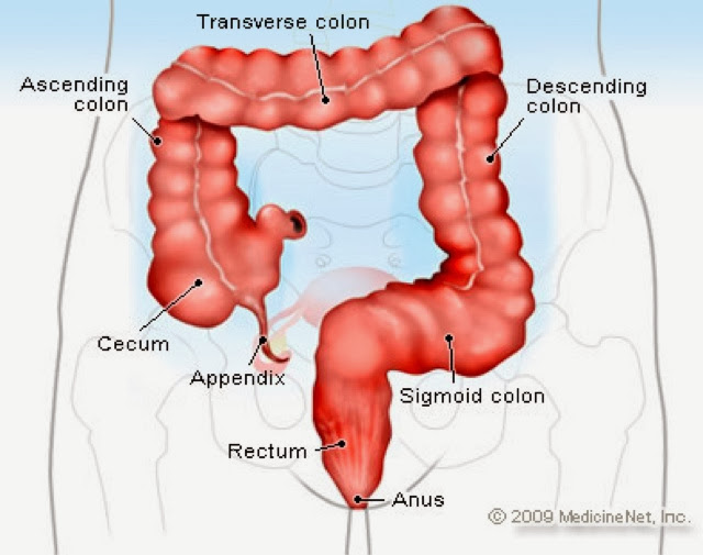
16. Appendix: sits at the junction of the small intestine and large intestine. It’s a thin tube about four inches long. The function of the appendix is not known.
17. Large Intestine(colon): it is larger in diameter but is much shorter in length than the small intestine. It has 4 main parts which are the ascending colon, transverse colon, descending colon and sigmoid colon(rectum). The large intestine absorbs water and salts that were used in the digestive process. It also has E.Coli bacteria that live in it. The E.Coli has four things that they do which are, slow the movement through the colon which allows time for water to be reabsorbed, they eat the wastes and produce useful things that we new to survive like vitamin K and amino acids, they also produce growth factor and the produce waste of there own (methane gas). By the end of the large intestines the waste are transformed into feces. If the waste move through the intestine too quickly it cannot absorb enough water resulting in diarrhea, if it goes to slow it will absorbed too much water resulting in constipation.
18. Rectum: Is the terminal portion of the large intestine, extending from the sigmoid colon to the anal canal. It acts as a temporary storage site for feces.
19. Anus: is the last part of the digestive tract. The anus is where the feces leaves the body.
The 3 Hormones that control digestion!
Gastrin: when food is present in the stomach gastrin is released and this causes the cells of the stomach mucosa to release gastric juices.
Secretin: is released when there is acidic chyme in the duodenum, this hormone causes the pancreatic juices to be released.
Cholecystokinin (CCK): is released when fats and proteins enter the duodenum, it is released into the blood stream and I causes the gall bladder to secrete bile and the pancreas to release it's pancreatic juices with digestive enzymes. These juices are sent to the duodenum where the enzymes can begin the digestion of lipids and proteins.
And that is everything I know about the digestive system!
Subscribe to:
Comments (Atom)